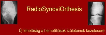
Dr. Gál János gondolatai
Tisztelt Szerkesztők, Tisztelt Hemofíliás Betegek!
Szeretettel üdvözlöm valamennyiüket.
Mivel időnként újabb és újabb becsületembe gázoló személyemet érintő kijelentéseket hallok vissza, ezért „hemofíliához nem értő doktorként” a WOLD FEDERATION OF HEMOPHILIA, vagyis a HEMOFÍLIA VILÁGSZÖVETSÉG módszertani útmutatójának áttekintését ajánlom mindenkinek.
Orvosoknak, betegegyesületeknek és azok elnökeinek is.
Egy hemofília betegegyesület vezetőjétől és mindenkitől aki bármely vonatkozásban hemofíliás betegekkel foglakozik joggal el lehet várni, hogy ezt ismerje és képviselje. Ha mégsem képviseli akkor vagy nagyon tudatlan, vagy szándékosan szembe megy a WOLD FEDERATION OF HEMOPHILIA hatályos ajánlásaival.
Egy ilyen ajánlást persze lehet kritika tárgyává tenni, de csak szakmai alapon. Érvek és ellenérvek ütköztetésével.
Viszont olyanokat terjeszteni „igazságként” aminek homlokegyenest az ellenkezője szerepel a VILÁGSZÖVETSÉG módszertani útmutatójában és közben arra hivatkozni, hogy „a szakirodalom nem rendelkezik ismeretekkel az eljárás hosszú távú hatásait illetően ” meg hogy „A nyugati orvostudomány ezt mondja meg azt mondja…” meg hogy „ Minden komoly szakember, nagyon határozottan leszögezte, hogy ezt az eljárást csak nagyon kevés esetben lehet, illetve érdemes alkalmazni „ nos efféléket már nem lenne szabad.
Ez esetben az illető megszegi a tízparancsolat azon pontját, hogy „Ne hazudj,mások becsületében kárt ne tégy!”, vagy ahogy az evangélikusok mondják: „Ne tégy felebarátod ellen hamis tanúbizonyságot!”. Héberül: „לֹא־תַעֲנֶה בְרֵעֲךָ עֵד שָׁקֶר׃” (a böngésző miatt hibásan jelenhet meg szerk.*)
Mindenkinek úgy ahogy számára ismertebb és hozzá közelebb áll.
Szeretném felhívni figyelmüket a következő site-ra:
http://www.wfh.org/2/docs/Publications/Diagnosis_and_Treatment/Guidelines_Mng_Hemophilia.pdf
It olvasható a GUDELINES FOR THE MANAGEMENT OF HEMOPHILIA
( Published by the World Federation of Hemophilia )
Témánkhoz a „Musculoskeletal Complications of Hemophilia” fejezet áll legközelebb, ebből ajánlom figyelmükbe a következőket:
MUSCULOSKELETAL COMPLICATIONS OF HEMOPHILIA
Introduction
The most common sites of bleeding in a person with hemophilia are the joints and muscles of the extremities. Depending on the severity of the disease, based on factor levels, the bleeding episodes are frequent and spontaneous in severe hemophilia (< 1%) or occur mostly after minor trauma in moderate hemophilia (1–5%). In the person with mild hemophilia (5–40%) bleeding usually occurs only with major trauma or surgery.
Common orthopedic complications of hemophilia are described below.
Acute Hemarthrosis
- In the child with severe hemophilia, the first spontaneous hemarthrosis typically occurs before two years of age, but may occur later.
- If inadequately treated, repeated bleeding will lead to progressive deterioration of the joint and muscles.
- This will lead to severe loss of function due to joint deformity, loss of motion, muscle atrophy, and contractures within the first one to two decades of life.
- The origin of the bleeding is the synovium. This is a very delicate and highly vascular tissue that lines and lubricates the joint space.
- Very early bleeding in joints may be recognized by the person experiencing it as a tingling sensation and tightness within the joint. This “aura” precedes the actual occurrence of the clinical features of acute hemarthrosis – pain, swelling, and limitation of motion.
- Once blood fills the joint cavity, the joint will appear swollen and feel warm and tender. This will cause the joint to seek the most comfortable position, which is flexion. Any attempt to change this position causes more pain, thus limiting motion. Secondary muscle spasm follows as the patient tries to prevent any motion.
- The goal of treatment of acute hemarthrosis is to stop the bleeding as soon as possible. Ideally, this should occur when the person recognizes the “aura”.
- The most important initial step in the management of the acute hemarthrosis is factor replacement as soon as possible at a level sufficiently high to stop the bleeding. (See Table 1, page 45)
- The most effective method of providing immediate factor replacement is to have a home therapy program in place that allows the informed patient with hemophilia (or his family members) to give the factor at the appropriate time. Joint bleeding that does not respond within 12-24 hours should be evaluated by a healthcare provider.
- Other measures to help control the bleeding and provide pain relief are as follows:
- Rest in the position of comfort.
- Immobilization (partial and temporary) with splints, pillows, slings, and crutches depending on the joint affected.
- Ice packs can be applied immediately and continued for at least the first 12 hours. Ice should not be in direct contact with the skin.
- Elevation of the affected joint.
- Tolerable pressure bandage can be used.
- Use of narcotics as analgesics should be carefully monitored, but preferably avoided because of the chronic nature of the bleeding episodes and the risks of addiction.
- Non-steroidal anti-inflammatory drugs (NSAIDS) and medications containing ASA are contraindicated during the acute bleeding episode. However, certain COX-2 inhibitor NSAIDs may be used judiciously.
- Physiotherapy must be stressed as an active part of the management of acute joint bleeding episodes.
- As soon as the pain and swelling begin to subside, the patient should attempt to change the position of the affected joint from a position of comfort to a position of function.
- This means gradually decreasing the flexion of the joint and striving for complete extension.
- This should include gentle, passive stretching and, more importantly, active muscle contractions to gain extension.
- The sooner the joint is in a position of function, the sooner active muscle control is instituted and this will in turn prevent muscle atrophy and loss of joint motion.
Aspiration
With an acute hemarthrosis, aspiration (removal of the blood from a joint) may be considered under certain circumstances. Once there is a large accumulation of blood in a joint, the early removal of the blood should result in a rapid relief of pain and theoretically reduce the damaging effects of the blood on the articular cartilage. Joint aspiration is usually not practical, however, because ideally it should be done very early following a bleeding episode (< 12 hours) and must be done in a medical facility by a physician.
- • Situations when joint aspiration may be considered include:
- A hemarthrosis that has not responded to factor replacement within 48–72 hours;
- Pain and swelling out of proportion with bleeding alone, in which circumstances a septic joint must be ruled out.
- No aspiration should be done when there is overlying skin infection.
- The presence of inhibitors should also be considered as a reason for persistent bleeding in the face of adequate factor replacement and must be ruled out before aspiration is attempted.
- When aspiration is performed, it should be done under factor levels of at least 30–50% for 48–72 hours. Joint aspiration should not be done in circumstances where such factor replacement is not available.
- A large bore needle, at least 16-gauge, should be used.
- The joint should be completely immobilized for one hour after the aspiration.
Muscle Hematomas
The sites of soft tissue and muscle bleeding that need immediate management are those affecting the flexor muscle groups in the arms and legs. The most critical sites of bleeding are those that have a risk of compromising the neurovascular function. These include:
- The iliopsoas muscle which may cause femoral nerve palsy;
- The gastrocnemius muscle which causes posterior tibial nerve injury and muscle contracture leading to an equinus deformity; and
- The flexor group of forearm muscles causing a Volkmann’s ischemic contracture.
Management of these bleeds include the following:
- These muscle bleeds require thorough clinical evaluation and monitoring.
- Factor replacement should be initiated immediately.
- Severe bleeds in these critical sites may require higher levels of factor replacement and for longer duration. (see Table 1, page 45)
- Other measures as discussed above for the acute joint bleed, such as elevation of the affected limb and physiotherapy, should also be included in the management of the acute muscle bleed.
Chronic Hemarthrosis
Once a joint develops recurrent bleeding episodes (target joint), chronic changes occur. These changes affect all of the tissues within and surrounding the joint: synovium and cartilage, capsule and ligaments, bone and muscles.
- Chronic synovitis is usually seen in the first and second decades of life.
- The management of chronic hemophilic arthropathy depends on the stage at which it is seen.
Chronic synovitis
With repeated bleeding in a joint, the synovium becomes chronically inflamed and eventually hypertrophies, causing the joint to appear grossly swollen. This swelling is usually not tense, nor is it particularly painful. Muscle atrophy is often present while a relatively good range of motion of the joint is preserved. Diagnosis made by performing a detailed physical examination of the joint. The presence of
synovial hypertrophy may be confirmed by ultrasonography and MRI. Plain radiographs and particularly MRI will assist in defining the extent of articular changes. The goal of treatment is to control the synovitis and maintain good joint function. Options include:
- Daily exercise to improve muscle strength and maintain joint motion is of prime importance. Factor concentrate replacement ideally should be given with the frequency and dose levels sufficient to prevent recurrent bleeding.
- If concentrates are available in sufficient doses, short treatment courses (6-8 weeks) of secondary prophylaxis with intensive physiotherapy is beneficial.
- NSAIDs (COX-2 inhibitors).
- Intra-articular injection of a long-acting steroid.
Synovectomy
Synovectomy should be considered if a chronic synovitis persists with frequent recurrent bleeding not controlled by other means. Options for synovectomy include chemical or radioisotopic synoviorthesis and arthroscopic or open surgical synovectomy.
- Surgical synovectomy, whether open or arthroscopic, requires enormous resources from an experienced team, a dedicated hemophilia treatment centre, and a large supply of clotting factor. Surgical synovectomy is seldom necessary today and is only considered when other less invasive and equally effective procedures fail.
- Non-surgical synovectomy should be the procedure of choice for treating chronic hemophilic synovitis. Clearly, radioisotopic synovectomy using a pure beta emitter (phosphorus-32 or yttrium-90) is the most effective and least invasive. It has the fewest side effects and is done in a simple out-patient setting. It also requires minimal if any follow-up physiotherapy. Only a single dose of clotting factor is required with the single dose of isotope.
- If a radioisotope is not available then chemical synovectomy is an appropriate alternative. Either rifampicin or oxytetracycline chlorhydrate can be used. Chemical synovectomy requires weekly injections until the synovitis is controlled. These painful injections require medication, and a dose of clotting factor is required for each injection. The low cost of the chemical agent is offset by the need for multiple injections.
Chronic hemophilic arthropathy
This can develop any time from the second decade of life, sometimes earlier, depending on the severity of bleeding and its treatment. It is caused by a persistent chronic synovitis and recurrent hemarthroses resulting in irreversible damage to the joint cartilage.
- With advancing cartilage loss, a progressive arthritis condition develops along with secondary soft tissue contractures, muscle atrophy, and angular deformities.
- With advancing chronicity of the arthropathy, there is less swelling due to progressive fibrosis of the synovium and the capsule.
- Loss of motion is common with flexion contractures causing the most significant functional loss.
- Pain may or may not be present.
The radiographic features of chronic hemophilic arthropathy depend on the stage of involvement.
- Early changes will show soft tissue swelling, epiphyseal overgrowth, and osteoporosis.
- Cartilage space narrowing will vary from minimal to complete loss.
- Bony erosions and subchondral bone cysts will develop, causing irregular articular bony surfaces which can lead to angular deformities.
- Fibrous/bony ankylosis may be present.
The goal of treatment is to improve joint function and relieve pain. Treatment options for chronic hemophilic arthropathy will depend on:
- The stage of the condition;
- The patient’s symptoms; and
- The resources available.
Supervised physiotherapy is a very important part of management at this stage. Factor replacement is necessary if recurrent bleeding occurs during physiotherapy. Pain should be controlled with appropriate analgesics.
- Narcotics should be avoided when possible.
- NSAIDs (certain COX-2 inhibitors) may be used to relieve arthritic pain.
Conservative management techniques include:
- Serial casting to assist in correcting deformities; and
- Bracing and orthotics to support painful and unstable joints.
If these conservative measures fail to provide satisfactory relief of pain and improved function, surgical intervention may be considered. Adequate resources, including sufficient factor concentrates, must be available in order to proceed with any surgical procedure.
Surgical procedures, depending on the specific condition needing correction, may include:
- Radionucleotide synoviorthesis;
- Extra-articular soft tissue release to treat contractures;
- Arthroscopy to release intra-articular adhesions and correct impingement;
- Elbow synovectomy with radial head excision;
- Osteotomy to correct an angular deformity;
- Prosthetic joint replacement for severe disease involving a major joint (knee, hip, shoulder); and
- Arthrodesis of the ankle which provides excellent pain relief and correction of deformity with marked improvement in function.
(idézetek vége)
Azok számára, akik nem beszélnek angolul, vagy éppen az orvosi szakszöveg értelmezése nehéz néhány sorban összefoglalom a HEMOFÍLIA VILÁGSZÖVETSÉG Módszertani Útmutatója kiemelt részeinek magyar nyelvű összefoglalóját:
Synovectomia
- Meg kell fontolni a synovectomiát, ha ha más módon nem kontrollálható, gyakori vérzésekkel járó krónikus synovitis áll fenn. A synovectomia lehetőségei a kémiai és radioizotópos synovectomiát és az arthroscopos vagy nyílt sebészi synovectomiát foglalják magukban.
- Sebészi synovectomia, akár arthroscopos akár nyílt, rendkívüli erőforrásokat igényel, kezdve azzal hogy szükséges egy gyakorlott team, egy elkötelezett hemofília kezelési központ és nagy mennyiségben rendelkezésre álló alvadási faktor. Manapság a sebészi synovectomia ritkán szükséges és csak akkor jön szóba, ha más kevésbé invazív ugyanakkor ezzel azonos mértékben hatékony beavatkozás sikertelen.
- A krónikus hemofíliás synovitis kezelésére a „nem-sebészi” synovectomia a választandó módszer. Egyértelmű, hogy a radioizotópos synoverctomia a leghatékonyabb és legkevésbé invazív, legkevesebb mellékhatással járó beavatkozás.
Krónikus hemofíliás arthropathia
- Ezt a perzisztáló krónikus synovitis és visszatérő ízületi bevérzések okozzák, melyek az ízületi porc irreverzibilis károsodásához vezetnek.
…
- Az ízület előrehaladó arthropathiájával a tok synoviumának progresszív fibrozisa következményeként kevesebb a duzzanat.
- Gyakori a flexiós kontraktúrával járó mozgáskorlátozottság, mely a legnyilvánvalóbb funkcionális károsodás
- Fájdalom vagy van, vagy nincs
…
- A kezelés célja az ízületi funkció javítása és a fájdalom csökkentése
…
- ha a konzervatív beavatkozások nem biztosítanak kielégítő fájdalomcsökkenést és funkcionális állapotjavulást akkor sebészi beavatkozást kell fontolóra venni
- Attól függően, hogy milyen speciális állapotbeli korrekció szükséges az alábbiakat foglalják magukba:
- Radionuclid synoviorthesis
…
…
Egyértelmű tehát, hogy mind a „hemofíliás synovitis”, mind a „krónikus hemofíliás arthropathia” kezelésében a Világszövetség Módszertani Útmutatója a radioizotópos eljárásokat állítja előtérbe, azokat javasolja elsődlegesen fontolóra venni és a műtéti megoldásokat csak abban az esetben ajánlja, ha az előbbi sikertelen, vagy olyan célok elérésére, melyek másként nem megvalósíthatók (pl.: protetizálás, stb)
Tisztelt Olvasók!
A „Módszertani Útmutató” minden sora fontos információkat tartalmaz, ezért a teljes szöveg elolvasását javaslom. (http://www.wfh.org/2/docs/Publications/Diagnosis_and_Treatment/Guidelines_Mng_Hemophilia.pdf )
Amiket kiemeltem azok a vázizomrendszeri problémákra vannak kihegyezve, azon belül is elsődlegesen a radiosynoviorthesisre. Amennyiben az olvasóknak szakmai jellegű kérdései adódnak, úgy szerény tudásom legjavát adva örömmel válaszolok azokra.
Dr Gál János
