Leptospirosis
Dogs
Leptospirosis
Leptospirosis (also known as Weil's disease, canicola
fever, canefield fever, nanukayami fever or 7-day
fever) is a
bacterial
zoonotic disease caused by
spirochaetes of the
genus
Leptospira that affects
humans
and a wide range of animals, including mammals, birds, amphibians, and
reptiles. It was first described by
Adolph Weil in
1886 when
he reported an "acute infectious disease with
enlargement of spleen,
jaundice and
nephritis". The
pathogen, Leptospira-genus
bacteria was isolated in
1907 from
post mortem renal
tissue slice.
Though being recognised among the world's most common
zoonosis,
leptospirosis is a relatively rare bacterial
infection
in humans. The infection is commonly transmitted to humans by allowing
fresh
water that has been contaminated by animal
urine to come in
contact with unhealed breaks in the
skin,
eyes or with the
mucous membranes.
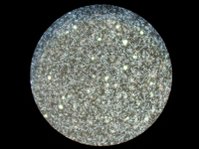 Leptospirose magnified 200 times with dark-field microscope
Except for tropic areas, leptospirosis cases have a relatively distinct
seasonality with most of them occurring August through September (in the
Northern
Causes
Leptospirosis is caused by a spirochaete bacterium called
leptospira
interrogans that has at least 4 different serovars of importance in the
United States causing disease (icterohaemorrhagiae, canicola, pomona,
grippotyphosa). There are other (less common) infectious strains. It should be
however noted that genetically different leptospira organisms may be identical
serologically and vice versa. Hence, an argument exists on the basis of strain
identification. The traditional serologic system is seemingfully more useful
from diagnostic and epidemiologic standpoint at the moment (which may change
with further development and spread of technologies like
PCR).
Leptospirosis is transmitted by the urine of an infected animal, and is
contagious as long as it is still moist. Rats, raccoons, possums, voles, skunks,
mice and even infected dogs may serve as hosts. Dogs may lick the urine of an
infected animal off the grass, or drink from an infected puddle. There have even
been reports of "house dogs" getting leptospirosis apparently from licking the
urine of infected mice that entered the house. There is a direct correlation
between the amount of rainfall and the incidence of leptospirosis.
Humans become infected through contact with water, food, or soil containing
urine from these infected animals. This may happen by swallowing contaminated
food or water or through skin contact. The disease is not known to be spread
from person to person and cases of bacteria dissemination in convalescence are
extremely rare in humans. Leptospirosis is common among watersport enthusiasts
in certain areas as prolonged immersion in water is known to promote the entry
of the bacteria.
Symptoms
In animals, the
incubation period (time of exposure to first
symptoms) is
anywhere from 2 to 20 days. One should strongly suspect leptospirosis and
include it as part of a
differential diagnosis if the whites of the dog's eyes appear
jaundiced
(even slightly yellow), but the absence of jaundice does not rule out
leptospirosis, and its presence could indicate
hepatitis
or liver pathology other rather than leptospirosis.
Vomiting, failure
to eat or drink, reduced urine output, unusually dark or brown urine,
lethargy,
and other such symptoms are also indications of the disease.
In humans, leptospiral infection causes a wide range of
symptoms, and
some infected persons may have no symptoms at all. Because of the wide range of
symptoms the infection is often
wrongly
diagnosed. This leads to a lower registered number of cases than there
really are. Symptoms of leptospirosis include high
fever, severe
headache,
chills, muscle aches, and
vomiting, and may
include
jaundice, red eyes,
abdominal pain,
diarrhea,
and/or a rash. The
symptoms in humans appear after 4-14 day incubation period.
Complications
Complications include
meningitis,
respiratory distress and renal interstitial tubular necrosis, which results in
renal
failure and often
liver
failure (this severe form of the disease is known as Weil's disease).
Cardiovascular problems are also possible. Approximately 5-50% of severe
leptospirosis cases are fatal, however, such cases only constitute about 10% of
all registered incidents.
The natural course of leptospirosis falls into 2 distinct phases, septicemic
and immune. During a brief period of 1-3 days between the 2 phases, the patient
shows some improvement.
First stage: This stage is called the septicemic or leptospiremic stage
because the organism may be isolated from blood cultures, cerebrospinal fluid (CSF),
and most tissues.
During this stage, which lasts about 4-7 days, the patient develops a
nonspecific flulike illness of varying severity.
It is characterized by fever, chills, weakness, and myalgias, primarily
affecting the calves, back, and abdomen.
Other symptoms are sore throat, cough, chest pain, hemoptysis, rash, frontal
headache, photophobia, mental confusion, and other symptoms of meningitis.
Because of the abrupt nature of the onset, the patient often can tell exactly
when the symptoms started.
During the 1-3 day period of improvement that follows the first stage, the
temperature curve drops and the patient may become afebrile and relatively
asymptomatic. The fever then recurs, indicating the onset of the second stage
when clinical or subclinical meningitis appears.
Second stage: This stage is called the immune or leptospiruric stage because
circulating antibodies may be detected or the organism may be isolated from
urine; it may not be recoverable from blood or CSF.
This stage occurs as a consequence of the body's immunologic response to
infection and lasts 0-30 days or more.
Disease referable to specific organs is seen. These organs include the
meninges, liver, eyes, and kidney.
Nonspecific symptoms, such as fever and myalgia, may be less severe than in
the first stage and last a few days to a few weeks.
Many patients (77%) experience headache that is intense and poorly controlled
by analgesics; this often heralds the onset of meningitis.
Anicteric disease: Aseptic meningitis is the most important clinical syndrome
observed in the immune anicteric stage.
Meningeal symptoms develop in 50% of patients. Cranial nerve palsies,
encephalitis, and changes in consciousness are less common. Mild delirium also
may be seen.
Symptoms may be nonspecific, and a viral etiology may be suspected.
Meningitis usually lasts a few days, but occasionally it can last 1-2
weeks.
Death is extremely rare in the anicteric cases.
Icteric disease: Leptospires may be isolated from the blood for 24-48 hours
after jaundice appears. Abdominal pain with diarrhea or constipation (30%),
hepatosplenomegaly, nausea, vomiting, and anorexia also are seen.
Uveitis (2-10%) can develop early or late in the disease and has been
reported to occur as late as 1 year after initial illness. Iridocyclitis and
chorioretinitis are other late complications that may persist for years. These
symptoms first manifest 3 weeks to 1 month after exposure. Subconjunctival
hemorrhage is the most common ocular complication of leptospirosis, occurring in
as many as 92% of patients. Leptospires may be present in the aqueous humor.
Renal symptoms such as azotemia, pyuria, hematuria, proteinuria, and oliguria
are seen in 50% of patients with leptospirosis. Leptospires may be present in
the kidney.
Pulmonary manifestations occur in 20-70% of patients.
Adenopathy, rashes, and muscular pain also are seen.
Clinical syndromes are not specific to the serotype, although some
manifestations may be seen more commonly with some serotypes.
Often, the serovar helps determine some of the more characteristic clinical
manifestations, but any leptospiral serovar can lead to the signs and symptoms
seen with this disease. For example, jaundice is seen in 83% of patients with L
icterohaemorrhagiae infection and in 30% of patients infected with L pomona. A
characteristic pretibial erythematous rash is seen in patients with L autumnalis
infection. Similarly, GI symptoms predominate in patients infected with L
grippotyphosa. Aseptic meningitis commonly occurs in those infected with L
pomona or L canicola.
Weil syndrome.
This severe form of leptospirosis primarily manifests as profound jaundice,
renal dysfunction, hepatic necrosis, pulmonary dysfunction, and hemorrhagic
diathesis.
It occurs at the end of the first stage and peaks in the second stage, but
the patient's condition can deteriorate suddenly at any time. Often the
transition between the stages is obscured.
o Fever may be marked during the second stage.
o Criteria to determine who will develop Weil disease are not well defined.
o Pulmonary manifestations include cough, dyspnea, chest pain, bloodstained
sputum, hemoptysis, and respiratory failure.
o Vascular and renal dysfunctions accompanied by jaundice develop 4-9 days
after onset of disease, and the jaundice may persist for weeks.
o Patients with severe jaundice are more likely to develop renal failure,
hemorrhage, and cardiovascular collapse. Hepatomegaly and tenderness in the
right upper quadrant may be present.
o Oliguric or anuric acute tubular necrosis may occur during the second week
due to hypovolemia and decreased renal perfusion.
o Multi-organ failure, rhabdomyolysis, adult respiratory distress syndrome,
hemolysis, splenomegaly (20%), congestive heart failure, myocarditis, and
pericarditis also may occur. o Weil syndrome carries a mortality rate of 5-10%.
The most severe cases of Weil syndrome, with hepatorenal involvement and
jaundice, carry a case-fatality rate of 20-40%. Mortality rate is usually higher
for older patients.
Leptospirosis may present with a macular or maculopapular rash, abdominal pain
mimicking acute appendicitis, or generalized enlargement of lymphoid glands
resembling infectious mononucleosis. It also may present as aseptic meningitis,
encephalitis, or fever of unknown origin.
Leptospirosis should be considered when a patient has a flulike disease with
aseptic meningitis or disproportionately severe myalgia.
Diagnostics
On infection the
microorganism can be found in
blood for the
first 7 to 10 days (invoking serologicaly identifiable reactions) and then
moving to the kidneys. After 7 to 10 days the microorganism can be found in
fresh urine. Hence, early diagnostic efforts include testing a serum or blood
sample serologically with a panel of different strains. It is also possible to
culture the microorganism from blood, serum, fresh urine and possibly fresh
kidney biopsy. Kidney function tests (Blood
Urea Nitrogen and
creatinine)
as well as blood tests for liver ferments are performed. The later reveal a
moderate elevation of transaminases.
Diagnosis
of leptospirosis is confirmed with tests such as
Enzyme-Linked Immunosorbent Assay (ELISA) and
PCR. It should be noted that serological testing is laborious and expensive,
thus underused in developing countries.
Differential diagnosis list for leptospirosis is very large due to diverse
symptomatics. For forms with middle to high severity, the list includes
dengue and
other hemorrhagic
fevers,
hepatitis of various
etiologies,
viral
meningitis,
malaria and
typhoid fever. Light forms should be distinguished from
influenza
and other related viral diseases. Specific tests are a must for proper diagnosis
of leptospirosis. Under circumstances of limited access (e.g., developing
countries) to specific diagnostic means, close attention must be paid to
anamnesis of the patient. Factors like certain dwelling areas, seasonality,
contact with stagnant water (swimming, working on flooded meadows, etc) and|or
rodents in the medical history support the leptospirosis hypothesis and serve
indictaions for specific tests (if available).
Treatment
Leptospirosis treatment is a relatively complicated process comprising two
main components - suppressing the causative agent and fighting possible
complications.
Aetiotropic drugs are
antibiotics, such as
doxycycline,
penicillin,
ampicillin,
and
amoxicillin (doxycycline can also be used as a
prophylaxis). There are no human
vaccines;
animal vaccines are only for a few strains, and are only effective for a few
months. Human
therapeutic dosage of drugs is as follows: doxycycline 100 mg orally every
12 hours for 1 week or penicillin 1-1.5 MU every 4 hours for 1 week. Doxycycline
200-250 mg once a week is administered as a
prophylaxis.
Supportive therapy measures (esp. in severe cases) include
detoxication and normalization of the
hydro-electrolytic balance. Glucose and salt solution infusions may be
administered;
dialysis is used in serious cases. Elevations of serum potassium are common
and if the potassium level gets too high special measures must be taken. Serum
phosphorus levels may likewise increase to unacceptable levels due to renal
failure. Treatment for hyperphosphatemia consists of treating the underlying
disease,
dialysis where appropriate, or oral administration of
calcium carbonate, but not without first checking the serum calcium levels
(these two levels are related).
Corticosteroids administraion in gradually reduced doses (e.g.,
prednisolone starting from 30-60 mg) during 7-10 days is recommended by some
specialists in cases of severe haemorrhagic effects.
Improper treatment greatly reduces the survival rate. A patient with
leptospirosis SHOULD be treated at a specialized medical institution and MUST
remain hospitalized untill proper resolution of organ(s) failure and clinical
infection.
Research
In a study of 38 dogs diagnosed and properly treated for leptospirosis
published in the February 2000 issue of the Journal of the American
Veterinary Association, the survival rate for the dialysis patients was
slightly higher than the ones not put on dialysis, but both were in the 85%
range (plus or minus). Of the dogs in this study that did not die, most
recovered adequate kidney function, although one had chronic renal problems.
External links
-
[1] U.S. Disease Control and Prevention Center page on Leptospirosis
-
[2] www.leptonet.net - the Leptospirosis information portal
-
[3] International Leptospirosis Society page
Home | Up | Legg-Calvé-Perthes Syndrome | Leptospirosis | Luxating Patella | Lymphoma
Dogs, made by MultiMedia | Free content and software
This guide is licensed under the GNU
Free Documentation License. It uses material from the Wikipedia.
|