Mastocytoma
Dogs
Mastocytoma
A Mastocytoma is an accumulation or
nodule
of
mast cells that resembles a tumor. In dogs and cats this collection
of mast cells is actually a mast cell tumor. A mast cell
originates from the
bone marrow and is normally found throughout the
connective tissue of the body. It is associated with
allergic reactions because it releases
histamine. A mast cell tumor is a common
malignant tumor of the skin in older dogs and cats.
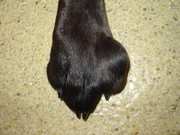 Mast cell tumor of the paw
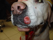 Mast cell tumor on the lip
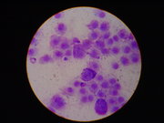 Mast cell tumor cytology
Commonly affected breeds
Symptoms
Most mast cell tumors are small, raised lumps on the skin. Some are hairless,
ulcerated, or
itchy. They are usually solitary. In rare cases a highly malignant tumor is
present, and symptoms may include loss of appetite,
vomiting,
diarrhea,
and anemia. The
presence of these symptoms usually indicates
mastocytosis, which is the spread of mast cells throughout the body. Release
of a large amount of histamine at one time can result in ulceration of the
stomach and
duodenum,
or
disseminated intravascular coagulation.
Diagnosis
A
needle aspiration biopsy of the tumor will show a large number of mast
cells. This is sufficient to make the diagnosis of a mast cell tumor. However, a
surgical biopsy
is required to find the
grade of the tumor. The grade depends on how well the mast cells are
differentiated, from grade I to grade III. The disease is also staged.
- Stage I - a single skin tumor with no spread to
lymph
nodes
- Stage II - a single skin tumor with spread to lymph nodes in the
surrounding area
- Stage III - multiple skin tumors or a large tumor invading deep to the
skin with or without lymph node involvement
- Stage IV - a tumor with
metastasis to the
spleen,
liver,
bone
marrow, or with the presence of mast cells in the blood
X-rays,
ultrasound,
or lymph node, bone marrow, or organ biopsies may be necessary to stage the
disease.
Treatment and prognosis
Removal of the mast cell tumor through surgery is the treatment of choice.
Antihistamines such as
diphenhydramine are given prior to surgery to protect against the effects of
histamine released from the tumor. Wide margins (two to three centimeters) are
required because of the tendency for the tumor cells to be spread out around the
tumor. If complete removal is not possible due to the size or location,
additional treatment such as
radiation therapy or
chemotherapy may be necessary.
Prednisone
is often used to shrink the remaining tumor portion.
H2 blockers
such as
cimetidine protect against stomach damage from histamine.
Mast cell tumors that are grade I or II that can be completely removed have a
good
prognosis. Any mast cell tumor found in the
gastrointestinal tract,
prepuce, paw,
or around the anus has a guarded prognosis. Tumors that have spread to the lymph
nodes or other parts of the body have a poor prognosis. Any dog showing symptoms
of mastocytosis or with a grade III tumor has a poor prognosis. Boxers have a
better than average prognosis because of the relatively benign behavior of their
mast cell tumors.
Mast cell tumors in cats
Siamese cats are at an increased risk for mast cell tumors. Gastrointestinal
and splenic
involvement is more common in cats. Diagnosis and treatment are similar to the
dog. The prognosis for solitary skin tumors is good, but guarded for tumors in
other organs.
References
- Morrison, Wallace B. (1998). Cancer in Dogs and Cats (1st ed.).
Williams and Wilkins.
ISBN 0-683-06105-4
Home | Up | Malignant Histiocytosis | Mange | Masticatory Muscle Myositis | Mastocytoma | Megaesophagus | Molera
Dogs, made by MultiMedia | Free content and software
This guide is licensed under the GNU
Free Documentation License. It uses material from the Wikipedia.
|